The circulatory system, also called the cardiovascular system or the vascular system, is an organ system that permits blood to circulate and transport nutrients (such as amino acids and electrolytes), oxygen, carbon dioxide, hormones, and blood cells to and from the cells in the body to provide nourishment and help in fighting diseases, stabilize temperature and pH, and maintain homeostasis. The study of the blood flow is called hemodynamics. The study of the properties of the blood flow is called hemorheology.
The circulatory system is often seen to comprise both the cardiovascular system, which distributes blood, and the lymphatic system, which circulates lymph.[1] These are two separate systems. The passage of lymph for example takes a lot longer than that of blood.[2] Blood is a fluid consisting of plasma, red blood cells, white blood cells, and platelets that is circulated by the heart through the vertebrate vascular system, carrying oxygen and nutrients to and waste materials away from all body tissues. Lymph is essentially recycled excess blood plasma after it has been filtered from the interstitial fluid (between cells) and returned to the lymphatic system. The cardiovascular (from Latin words meaning 'heart' and 'vessel') system comprises the blood, heart, and blood vessels.[3] The lymph, lymph nodes, and lymph vessels form the lymphatic system, which returns filtered blood plasma from the interstitial fluid (between cells) as lymph.
While humans, as well as other vertebrates, have a closed cardiovascular system (meaning that the blood never leaves the network of arteries, veins and capillaries), some invertebrate groups have an open cardiovascular system. The lymphatic system, on the other hand, is an open system providing an accessory route for excess interstitial fluid to be returned to the blood.[4] The more primitive, diploblastic animal phyla lack circulatory systems.
Structure
Cardiovascular system

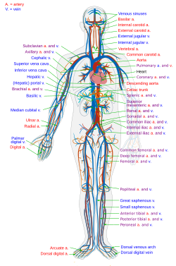
Depiction of the heart, major veins and arteries constructed from body scans.

Cross section of a human artery

Relative percentages of cardiac output delivered to major organ systems
The cardiovascular systems of humans are closed, meaning that the blood never leaves the network of blood vessels. In contrast, oxygen and nutrients diffuse across the blood vessel layers and enter interstitial fluid, which carries oxygen and nutrients to the target cells, and carbon dioxide and wastes in the opposite direction. The other component of the circulatory system, the lymphatic system, is open.
Arteries
See also: Arterial tree
Oxygenated blood enters the systemic circulation when leaving the left ventricle, through the aortic semilunar valve. The first part of the systemic circulation is the aorta,
a massive and thick-walled artery. The aorta arches and branches into
major arteries to the upper body before passing through the diaphragm,
where it branches further into arteries which supply the lower parts of
the body.Capillaries
Arteries branch into small passages called arterioles and then into the capillaries.[8] The capillaries merge to bring blood into the venous system.[9]Veins
After their passage through body tissues, capillaries merge once again into venules, which continue to merge into veins. The venous system finally coalesces into two major veins: the superior vena cava (roughly speaking draining the areas above the heart) and the inferior vena cava (roughly speaking from areas below the heart). These two great vessels empty into the right atrium of the heart.Coronary vessels
Main article: Coronary circulation
The heart itself is supplied with oxygen and nutrients through a small "loop" of the systemic circulation.Portal veins
The general rule is that arteries from the heart branch out into capillaries, which collect into veins leading back to the heart. Portal veins are a slight exception to this. In humans the only significant example is the hepatic portal vein which combines from capillaries around the gut where the blood absorbs the various products of digestion; rather than leading directly back to the heart, the hepatic portal vein branches into a second capillary system in the liver.Heart
Main article: Heart

View from the front
The coronary circulation system provides a blood supply to the heart muscle itself. The coronary circulation begins near the origin of the aorta by two arteries: the right coronary artery and the left coronary artery. After nourishing the heart muscle, blood returns through the coronary veins into the coronary sinus and from this one into the right atrium. Back flow of blood through its opening during atrial systole is prevented by the Thebesian valve. The smallest cardiac veins drain directly into the heart chambers.[7]
Lungs

The pulmonary circulation as it passes from the heart. Showing both the pulmonary artery and bronchial arteries.
Main article: Pulmonary circulation
The circulatory system of the lungs is the portion of the cardiovascular system in which oxygen-depleted blood is pumped away from the heart, via the pulmonary artery, to the lungs and returned, oxygenated, to the heart via the pulmonary vein.Oxygen deprived blood from the superior and inferior vena cava enters the right atrium of the heart and flows through the tricuspid valve (right atrioventricular valve) into the right ventricle, from which it is then pumped through the pulmonary semilunar valve into the pulmonary artery to the lungs. Gas exchange occurs in the lungs, whereby CO2 is released from the blood, and oxygen is absorbed. The pulmonary vein returns the now oxygen-rich blood to the left atrium.[7]
A separate system known as the bronchial circulation supplies blood to the tissue of the larger airways of the lung.
Systemic circulation
The systemic circulation is the circulation of the blood to all parts of the body except the lungs. Systemic circulation is the portion of the cardiovascular system which transports oxygenated blood away from the heart through the aorta from the left ventricle where the blood has been previously deposited from pulmonary circulation, to the rest of the body, and returns oxygen-depleted blood back to the heart.[7]Brain
Main article: Cerebral circulation
The brain has a dual blood supply that comes from arteries at its
front and back. These are called the "anterior" and "posterior"
circulation respectively. The anterior circulation arises from the internal carotid arteries and supplies the front of the brain. The posterior circulation arises from the vertebral arteries, and supplies the back of the brain and brainstem. The circulation from the front and the back join together (anastomise) at the Circle of Willis.Kidneys
The renal circulation receives around 20% of the cardiac output. It branches from the abdominal aorta and returns blood to the ascending vena cava. It is the blood supply to the kidneys, and contains many specialized blood vessels.Lymphatic system
The lymphatic system is part of the circulatory system. It is a network of lymphatic vessels and lymph capillaries, lymph nodes and organs, and lymphatic tissues and circulating lymph. One of its major functions is to carry the lymph, draining and returning interstitial fluid back towards the heart for return to the cardiovascular system, by emptying into the lymphatic ducts. Its other main function is in the immune system.Physiology

An animation of a typical human red blood cell cycle in the circulatory
system. This animation occurs at real time (20 seconds of cycle) and
shows the red blood cell deform as it enters capillaries, as well as
changing color as it alternates in states of oxygenation along the
circulatory system.
Main article: Blood § Oxygen transport
About 98.5% of the oxygen in a sample of arterial blood in a healthy human, breathing air at sea-level pressure, is chemically combined with hemoglobin
molecules. About 1.5% is physically dissolved in the other blood
liquids and not connected to hemoglobin. The hemoglobin molecule is the
primary transporter of oxygen in mammals and many other species.Development
Main article: Fetal circulation
The development of the circulatory system starts with vasculogenesis in the embryo.
The human arterial and venous systems develop from different areas in
the embryo. The arterial system develops mainly from the aortic arches,
six pairs of arches which develop on the upper part of the embryo. The
venous system arises from three bilateral veins during weeks 4 – 8 of embryogenesis. Fetal circulation begins within the 8th week of development. Fetal circulation does not include the lungs, which are bypassed via the truncus arteriosus. Before birth the fetus obtains oxygen (and nutrients) from the mother through the placenta and the umbilical cord.[10]Arterial development
Main article: Aortic arches
The human arterial system originates from the aortic arches and from the dorsal aortae starting from week 4 of embryonic life. The first and second aortic arches regress and forms only the maxillary arteries and stapedial arteries respectively. The arterial system itself arises from aortic arches 3, 4 and 6 (aortic arch 5 completely regresses).The dorsal aortae, present on the dorsal side of the embryo, are initially present on both sides of the embryo. They later fuse to form the basis for the aorta itself. Approximately thirty smaller arteries branch from this at the back and sides. These branches form the intercostal arteries, arteries of the arms and legs, lumbar arteries and the lateral sacral arteries. Branches to the sides of the aorta will form the definitive renal, suprarenal and gonadal arteries. Finally, branches at the front of the aorta consist of the vitelline arteries and umbilical arteries. The vitelline arteries form the celiac, superior and inferior mesenteric arteries of the gastrointestinal tract. After birth, the umbilical arteries will form the internal iliac arteries.
Venous development
The human venous system develops mainly from the vitelline veins, the umbilical veins and the cardinal veins, all of which empty into the sinus venosus.Clinical significance
Many diseases affect the circulatory system. This includes cardiovascular disease, affecting the cardiovascular system, and lymphatic disease affecting the lymphatic system. Cardiologists are medical professionals which specialise in the heart, and cardiothoracic surgeons specialise in operating on the heart and its surrounding areas. Vascular surgeons focus on other parts of the circulatory system.Cardiovascular disease
Main article: Cardiovascular disease
Diseases affecting the cardiovascular system are called cardiovascular disease.Many of these diseases are called "lifestyle diseases" because they develop over time and are related to a person's exercise habits, diet, whether they smoke, and other lifestyle choices a person makes. Atherosclerosis is the precursor to many of these diseases. It is where small atheromatous plaques build up in the walls of medium and large arteries. This may eventually grow or rupture to occlude the arteries. It is also a risk factor for acute coronary syndromes, which are diseases which are characterised by a sudden deficit of oxygenated blood to the heart tissue. Atherosclerosis is also associated with problems such as aneurysm formation or splitting ("dissection") of arteries.
Another major cardiovascular disease involves the creation of a clot, called a "thrombus". These can originate in veins or arteries. Deep venous thrombosis, which mostly occurs in the legs, is one cause of clots in the veins of the legs, particularly when a person has been stationary for a long time. These clots may embolise, meaning travel to another location in the body. The results of this may include pulmonary embolus, transient ischaemic attacks, or stroke.
Cardiovascular diseases may also be congenital in nature, such as heart defects or persistent fetal circulation, where the circulatory changes that are supposed to happen after birth do not. Not all congenital changes to the circulatory system are associated with diseases, a large number are anatomical variations.
Measurement techniques

Other more invasive means can also be used. A cannula or catheter inserted into an artery may be used to measure pulse pressure or pulmonary wedge pressures. Angiography, which involves injecting a dye into an artery to visualise an arterial tree, can be used in the heart (coronary angiography) or brain. At the same time as the arteries are visualised, blockages or narrowings may be fixed through the insertion of stents, and active bleeds may be managed by the insertion of coils. An MRI may be used to image arteries, called an MRI angiogram. For evaluation of the blood supply to the lungs a CT pulmonary angiogram may be used.
Ultrasound can also be used, particularly to identify the health of blood vessels, and a Doppler ultrasound of the carotid arteries or Doppler ultrasound of the lower limbs can be used to evaluate for narrowing of the carotid arteries or thrombus formation in the legs, respectively.
Surgery
| This section requires expansion. (March 2015) |
- Coronary artery bypass surgery
- Coronary stent used in angioplasty
- Vascular surgery
- Vein stripping
- Cosmetic procedures
No comments:
Post a Comment